Ptosis, Entropion & Surgical Repair: A Complete Guide to Eyelid Disorders
 Jun, 20 2026
Your eyelids do more than just blink. They act as a windshield wiper system, spreading tears across your eye to keep it moist and protected. But when that delicate machinery starts to fail, the consequences can range from minor irritation to permanent vision loss. You might notice one eyelid sitting lower than the other, or perhaps you feel like there is sand constantly grinding against your eyeball. These are not just cosmetic quirks; they are signs of structural failures known as eyelid disorders, specifically conditions like ptosis (drooping) and entropion (inward turning).
If you are over 60, these issues become statistically likely. Approximately 5% of adults in this age group experience some form of eyelid malposition. The good news? Modern oculoplastic surgery has refined techniques that offer high success rates, often restoring both function and appearance. This guide breaks down exactly what is happening to your eyes, how doctors diagnose the problem, and what surgical repairs actually involve.
Understanding Ptosis: The Drooping Eyelid
Ptosis is the medical term for a drooping upper eyelid. It occurs when the muscle responsible for lifting the lid-the levator palpebrae superioris-weakens or disconnects. Think of it like a window shade where the cord has snapped or the spring has lost its tension. The lid falls down, potentially covering part or all of your pupil.
This isn't always about aging, though that is the most common culprit. In older adults, natural degeneration stretches the tendon connecting the muscle to the eyelid bone. However, ptosis can also be congenital (present at birth), caused by nerve damage from diabetes or stroke, or even triggered by prolonged contact lens wear, which increases risk by approximately 30%. If your drooping happens suddenly-within hours or days-it requires immediate medical attention, as it could signal a serious neurological event like an aneurysm.
Doctors measure the severity of ptosis using the Margin Reflex Distance (MRD). A normal MRD is between 4mm and 5mm. If your measurement drops to 1-2mm, it’s considered mild. Between 2-3mm is moderate, and anything greater than 3mm is severe. Why does this number matter? Because the treatment depends entirely on how much "lift" your muscle still has. If your muscle is strong but the tendon is stretched, one surgery works. If the muscle itself is weak, a completely different procedure is required.
Understanding Entropion: The Inward Turn
While ptosis blocks your view from above, Entropion is a condition where the eyelid margin turns inward toward the eye. This forces your eyelashes and skin to rub directly against the cornea-the clear front surface of your eye. Imagine walking around with a piece of grit stuck under your eyelid, every single second. That is the reality for patients with untreated entropion.
The symptoms are unmistakable: redness, excessive tearing, mucus discharge, and a persistent sensation of foreign body presence. Left alone, this friction causes corneal abrasions, ulcers, and scarring that can lead to permanent sight loss. Entropion predominantly affects the lower eyelid, accounting for 97% of cases.
There are four main types, but two dominate clinical practice:
- Involutional Entropion: This is the age-related type, making up about 80% of cases in Western populations. As we age, the horizontal muscles of the eyelid loosen, allowing the lid to roll inward. The incidence jumps from 0.5% in people aged 50-60 to 2.5% in those over 80.
- Cicatricial Entropion: Caused by scarring on the inner surface of the eyelid. This scar tissue contracts and pulls the lid inward. Common causes include chronic blepharitis, ocular rosacea, chemical burns, or previous surgeries. Trachoma, a bacterial infection caused by Chlamydia trachomatis, remains a leading cause of cicatricial entropion in regions with limited sanitation.
Diagnosis and Pre-Surgical Assessment
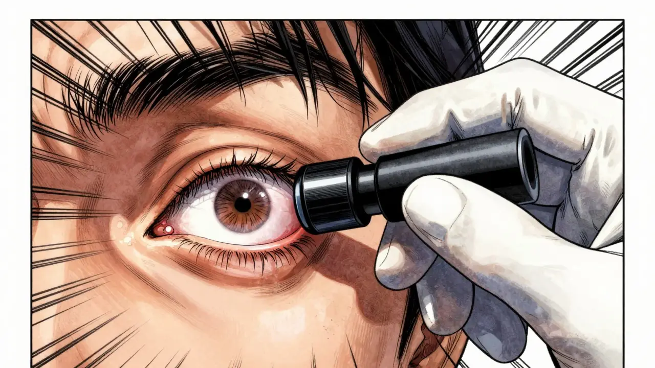
Before any knife touches skin, a precise diagnosis is critical. An ophthalmologist or oculoplastic surgeon will perform a slit-lamp examination to look for corneal damage. For ptosis, they will test the "levator function" by holding the brow down and asking you to look up. If the lid moves up significantly, the muscle is healthy. If it barely moves, the muscle is paralyzed or severely weak.
For mild ptosis, doctors may use a phenylephrine drop test. This medication temporarily tightens the Müller's muscle inside the lid. If the lid lifts nicely after the drop, you are a candidate for a less invasive procedure called Müller's muscle-conjunctival resection (MMCR). If it doesn’t move, you’ll need a more robust levator resection.
Recent advancements in preoperative imaging have improved planning accuracy by 30-40%. High-resolution photos and measurements help surgeons predict exactly how much tissue needs to be adjusted, reducing the guesswork that once led to higher revision rates.
| Feature | Ptosis | Entropion |
|---|---|---|
| Primary Symptom | Drooping upper lid, hooded eyes | Inward turning lid, gritty sensation |
| Affected Lid | Upper eyelid | Lower eyelid (97% of cases) |
| Risk to Vision | Blocked field of view, amblyopia in kids | Corneal ulceration, scarring, blindness |
| Most Common Cause | Aging (aponeurotic dehiscence) | Aging (involutional laxity) |
| Key Diagnostic Test | Levator function measurement | Slit-lamp exam for corneal abrasions |
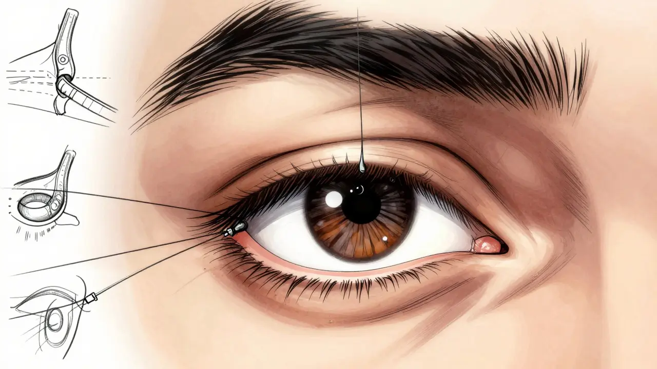
Surgical Repair Techniques for Ptosis
Surgery for ptosis is highly specialized. The goal is to restore the lid to a position that clears the pupil without making the eye look wide-eyed or unnatural. There are three primary surgical approaches, chosen based on your muscle strength and severity.
- Levator Resection: This is the gold standard for moderate to severe ptosis where the levator muscle still has good function (>4mm movement). The surgeon removes a small segment of the levator aponeurosis (tendon) and sutures it tighter. This shortens the path the muscle takes to lift the lid. Success rates hover between 85-95% for primary cases.
- Müller’s Muscle-Conjunctival Resection (MMCR): Ideal for mild ptosis (1-2mm droop) with a positive phenylephrine test. This procedure is performed from the inside of the eyelid, leaving no external scars. It’s faster, has less downtime, and carries a lower risk of asymmetry compared to levator surgery.
- Frontalis Sling Procedure: Reserved for severe cases where the levator muscle is essentially non-functional (<4mm movement), often due to congenital defects or nerve palsy. Since the eyelid muscle won’t work, surgeons borrow power from the forehead muscle (frontalis). A sling material connects the eyelid to the forehead. Now, when you raise your eyebrows, your eyelids open. This requires learning a new motor habit but restores functional vision.
A significant innovation introduced around 2018 is the use of adjustable sutures in ptosis surgery. Instead of fixing the height permanently during the operation, the surgeon leaves knots accessible outside the skin. After swelling goes down (usually 2-3 days post-op), the doctor can tweak the suture length to perfect the symmetry. This technique has reduced the need for revision surgery by approximately 25%.
Surgical Repair Techniques for Entropion
Fixing entropion is about restoring the mechanical balance of the eyelid. The lid must sit flat against the globe of the eye. Surgeons use different strategies depending on whether the cause is loose tissues (involutional) or tight scars (cicatricial).
- Tarsal Fracture Procedure: This is the most common fix for involutional entropion. The surgeon makes a small incision on the inside of the lid, creates a controlled break (fracture) in the tarsal plate (the stiff cartilage-like structure of the lid), and places sutures to anchor the lid in the correct outward position. It boasts a 90-95% success rate.
- Quickert Suture Technique: A temporary or semi-permanent solution. Silicone buttons are placed on the outer skin of the lid, connected by sutures that pull the lid outward. It’s less invasive and useful for elderly patients who cannot tolerate general anesthesia. However, the success rate is lower (60-70%) because the underlying laxity isn’t fully corrected.
- Tarsal Wedge Resection: Used primarily for cicatricial entropion. Since scar tissue is pulling the lid in, the surgeon removes a wedge-shaped piece of the tarsal plate to release the tension and straighten the lid margin.
Newer minimally invasive techniques using absorbable sutures have dramatically cut recovery time. Where patients once needed 4-6 weeks of healing, many now return to normal activities in 1-2 weeks with comparable long-term success.
Risks, Complications, and Recovery
No surgery is without risk, and eyelid procedures are no exception. Understanding these potential pitfalls helps set realistic expectations.
For ptosis surgery, the most common issues are overcorrection (lid too high, looking surprised) and undercorrection (lid still droopy). Overcorrection occurs in 5-10% of cases, while undercorrection happens in 3-8%. Asymmetry, where one eye looks different from the other, affects 5-15% of patients initially, though this often resolves as swelling subsides. Dry eye symptoms worsen in 10-20% of cases because the wider opening exposes more surface area to air.
Entropion surgery carries risks of recurrence (5-15%), especially if the underlying laxity was severe. Scarring (2-5%) and infection (1-3%) are rare but possible. If you have chronic blepharitis or ocular rosacea, these inflammatory conditions must be managed medically before and after surgery, or the recurrence rate skyrockets. Dr. Alison Bozung, OD, emphasizes that cicatricial ectropion/entropion related to systemic diseases like cutaneous T-cell lymphoma requires multidisciplinary care, not just local lid repair.
Recovery typically involves applying antibiotic ointment and cold compresses for the first 48 hours. Stitches are usually removed within 5-7 days. Most patients can read and drive within a week, though final aesthetic results take 3-6 months as subtle swelling disappears.
When to Seek Immediate Care
Not all eyelid changes require surgery immediately, but some are emergencies. You should seek urgent medical attention if:
- You experience sudden, rapid drooping of one or both eyelids.
- You have double vision, severe headache, or weakness in other parts of your face.
- Your eye becomes extremely painful, red, and sensitive to light (signs of corneal ulceration from entropion).
- You notice pus or significant discharge accompanied by fever.
For chronic, gradual changes, conservative management can provide temporary relief. Lubricating eye drops and ointments protect the cornea in entropion. Taping the eyelid upward at night can prevent exposure keratopathy. Warm compresses help manage associated blepharitis. However, these are band-aids. Structural problems like stretched tendons or rolled lids rarely fix themselves. Surgery remains the definitive cure for restoring both health and function.
Is ptosis surgery covered by insurance?
Yes, if the ptosis is functional. Insurance companies typically cover the procedure if the drooping eyelid obstructs your visual field. Your doctor will perform a visual field test to document the blockage. If the surgery is purely cosmetic (to improve appearance without vision obstruction), it is usually considered elective and not covered.
How long does the effect of ptosis surgery last?
In most cases, the results are permanent. However, because aging continues, the tissues may stretch again over many years. Revision surgery is sometimes needed, particularly if the initial correction was slight or if you had significant laxity beforehand. Adjustable sutures have helped reduce the frequency of these revisions.
Can entropion be fixed without surgery?
Temporary measures like taping the lid, using lubricating drops, or inserting a punctal plug can manage symptoms. The Quickert suture technique is a minimally invasive option that avoids major surgery but may need repeating. For true anatomical correction, especially in involutional entropion, surgery is the only reliable long-term solution to prevent corneal damage.
What is the difference between entropion and ectropion?
Entropion is when the eyelid turns inward, causing lashes to rub the eye. Ectropion is when the eyelid turns outward, exposing the inner lining and preventing tear drainage. Both are malpositions, but they present opposite symptoms: entropion causes pain and scratching, while ectropion causes dryness and watery eyes due to poor tear distribution.
Does contact lens wear cause ptosis?
Long-term contact lens wear is a risk factor. Studies suggest it increases the likelihood of developing acquired ptosis by approximately 30%, likely due to chronic stretching of the levator aponeurosis during insertion and removal. If you wear contacts and notice drooping, inform your ophthalmologist.